Morphology and Structure
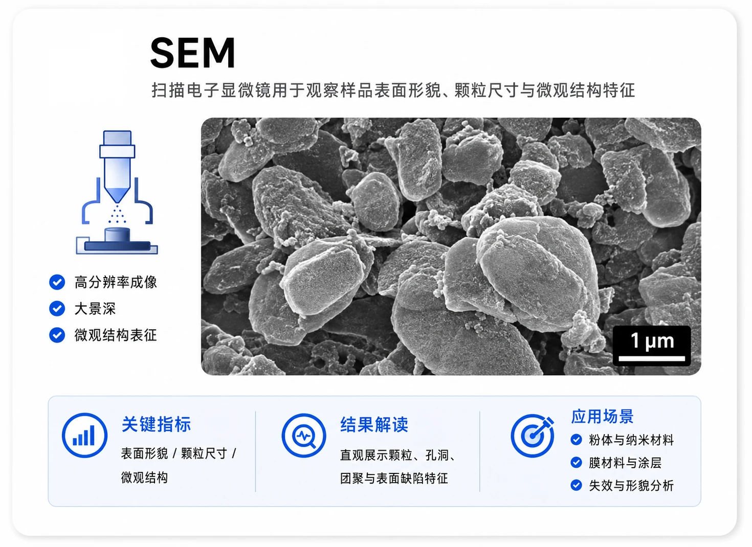
Scanning Electron Microscopy (SEM)
FE-SEM profile, multiple indicator, representative of FOV
Microimage, Cross-section sample, Reconstruct 3D, Crystal particle orientation and shape statistics.
FE-SEM profile, multiple indicator, representative of FOV
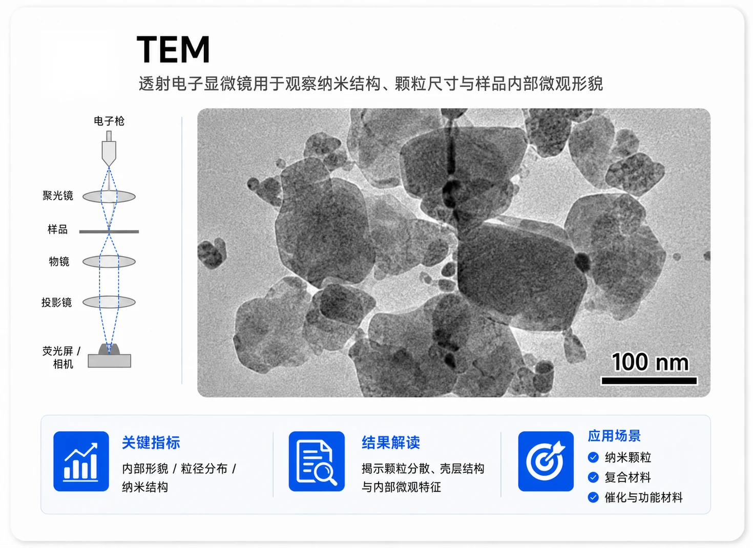
TEM/HRTEM chart, crystal stripes, size statistics; Based on regular TEM/HRTEM integrated project 500 yuan/sample reference.
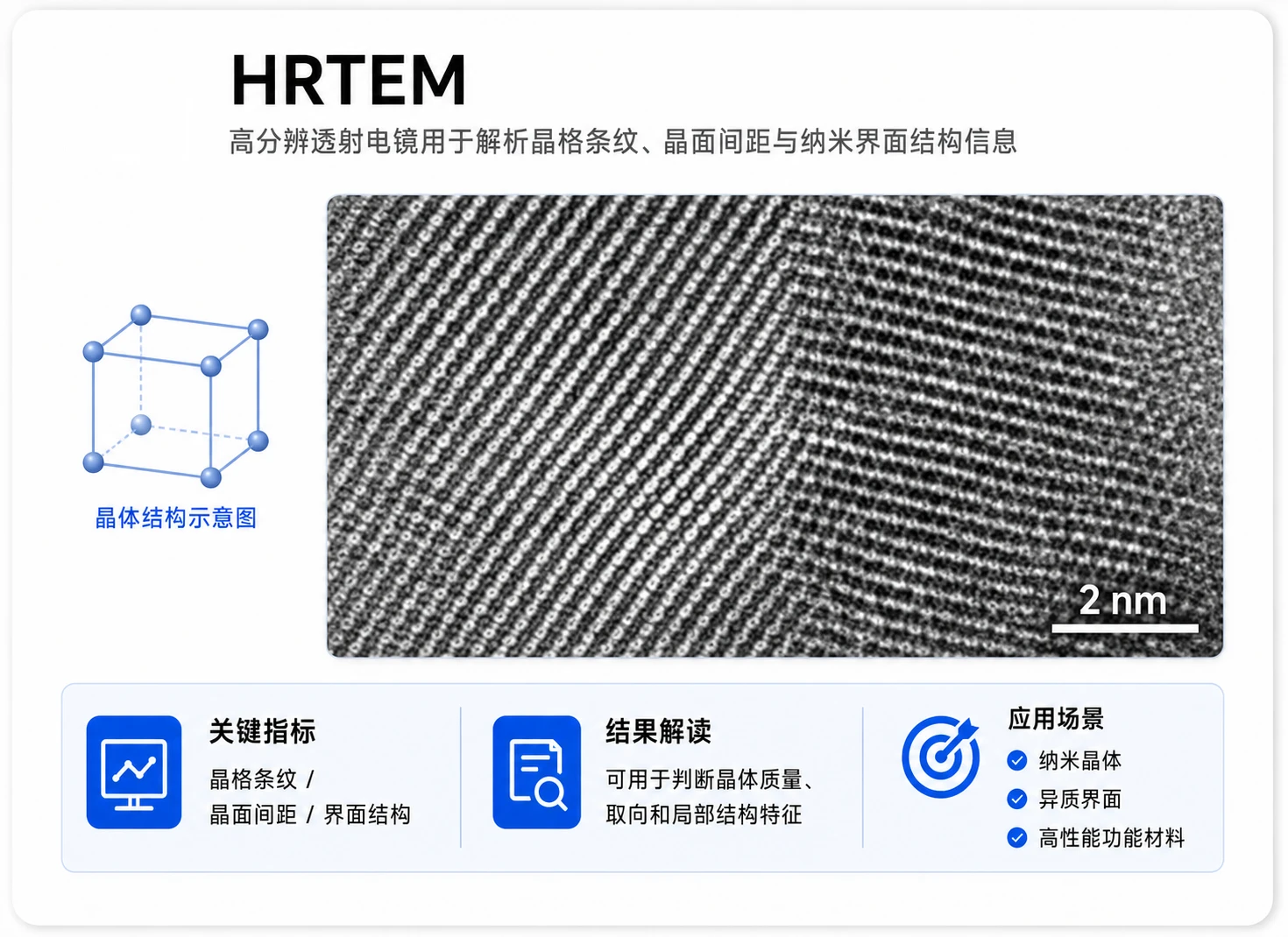
TEM/HRTEM chart, crystal stripes, size statistics; Based on TEM/HRTEM integrated project 500 yuan/sample.
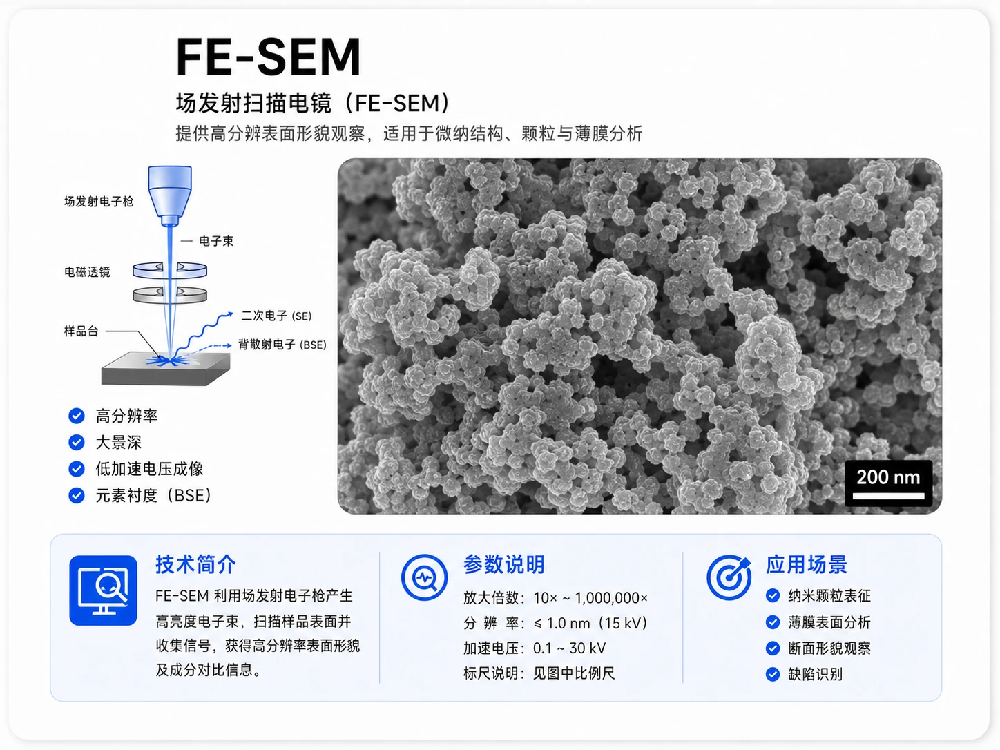
FE-SEM profile, multiple indicator, representative of FOV
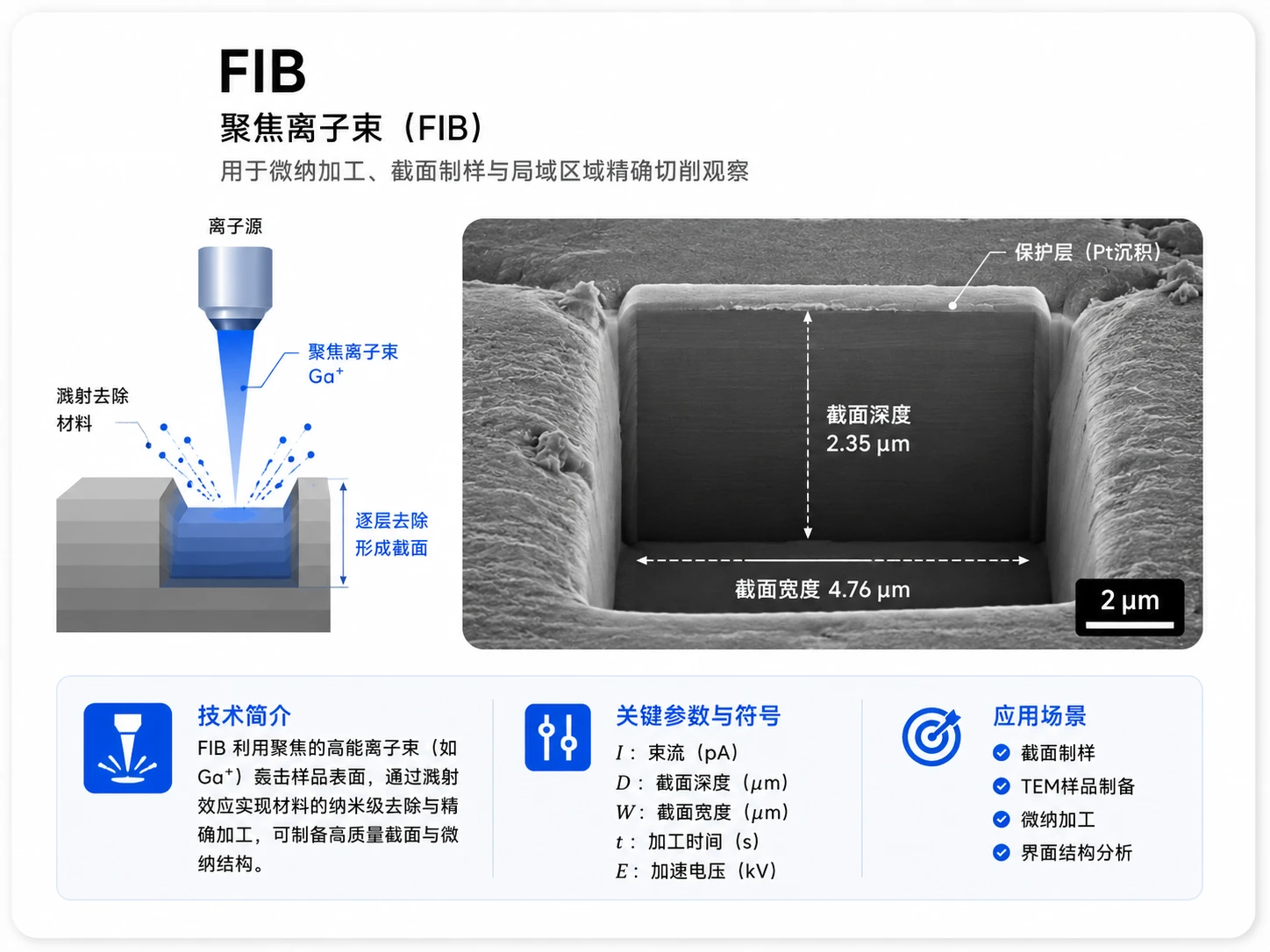
Cross-section, layer thick, three-dimensional reconstruction of data
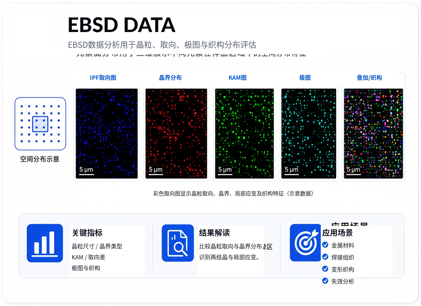
Crystal particles, orientation, polar maps, fabric distribution
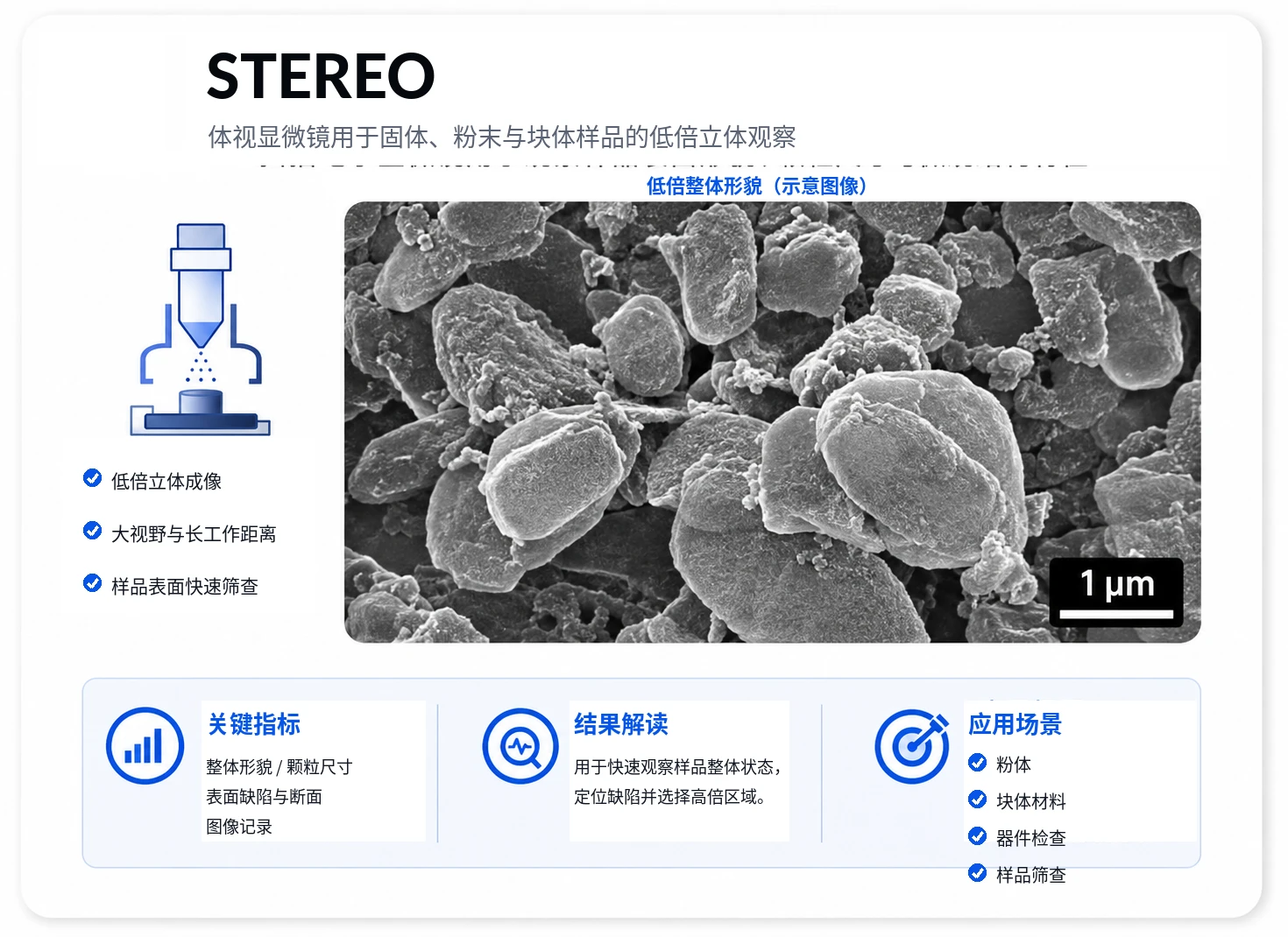
10-80X solid/powder/block observation
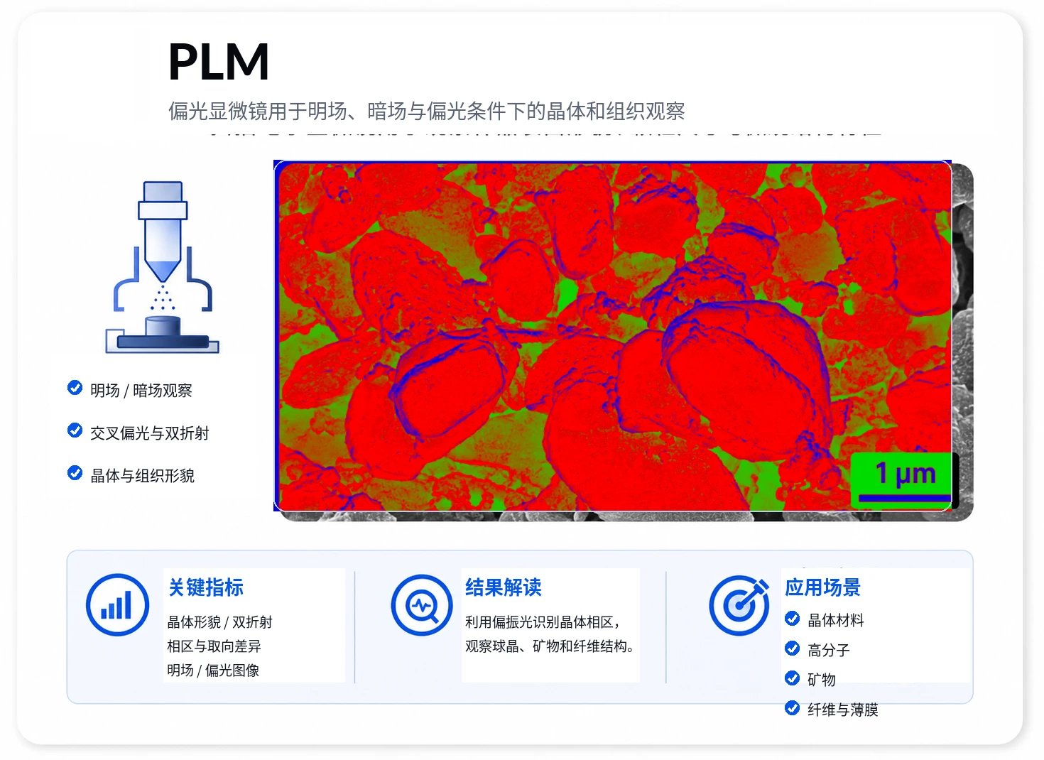
Open/dark/light observation
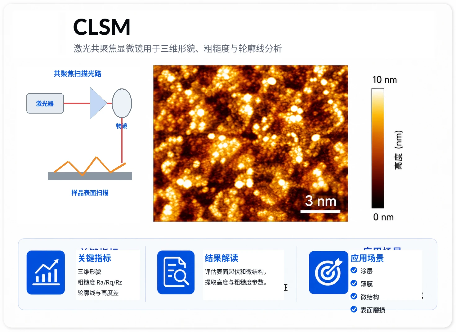
Three-dimensional shape, Roughness, contour
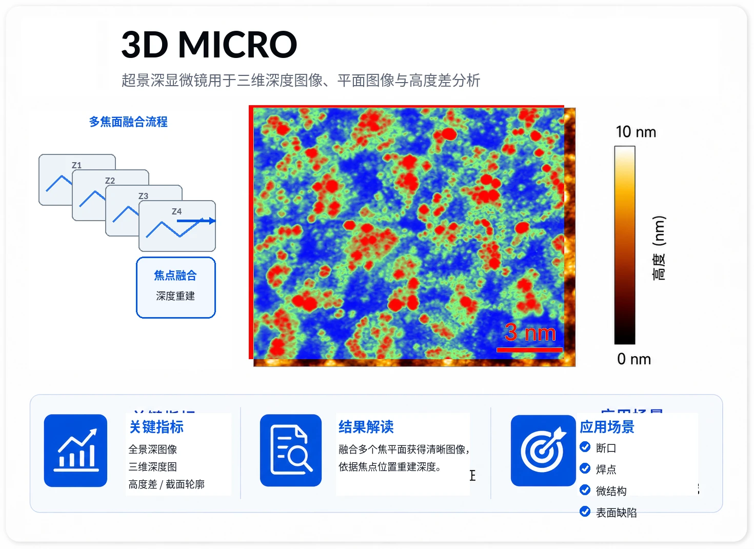
3D deep image and face image
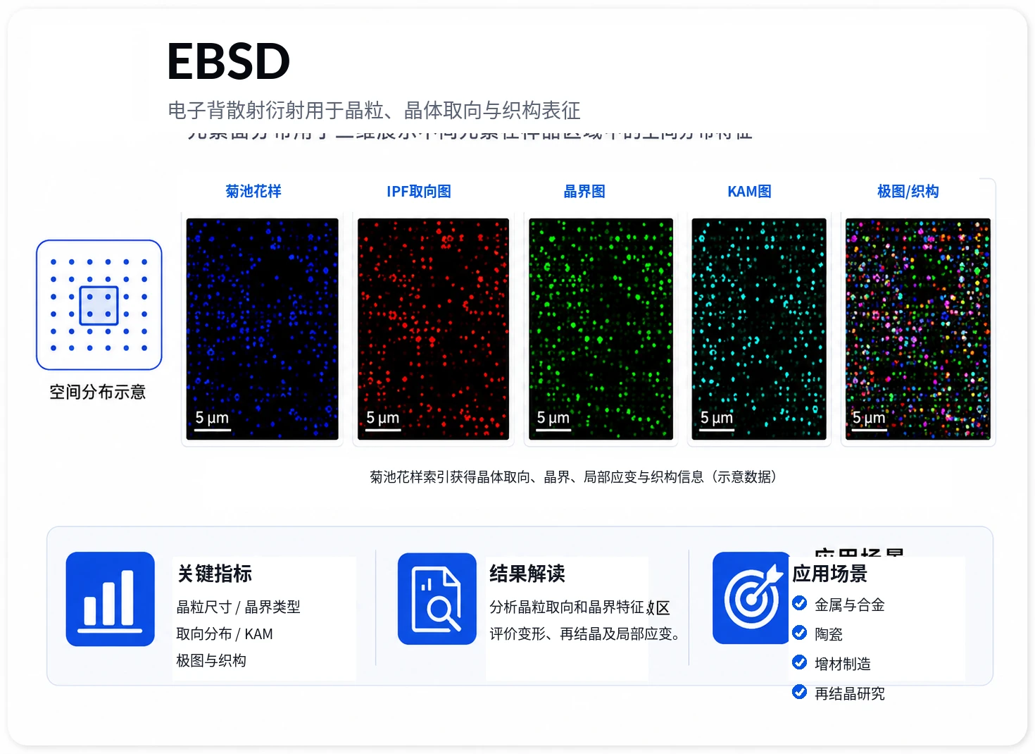
Crystal particles, direction, fabric
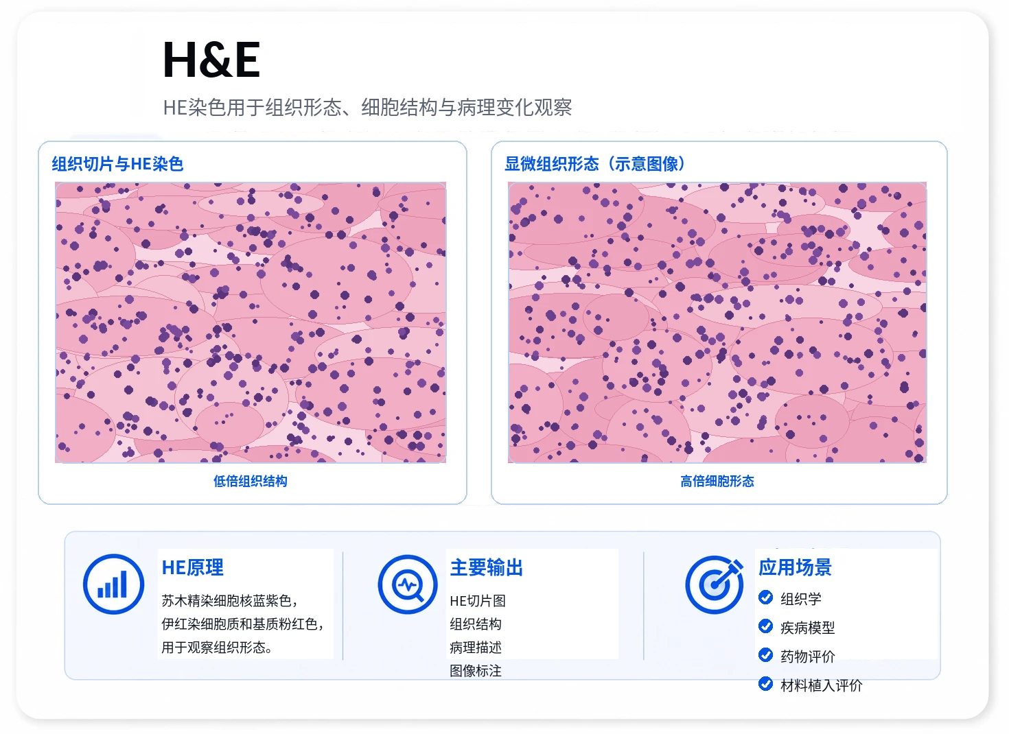
Organization dyed
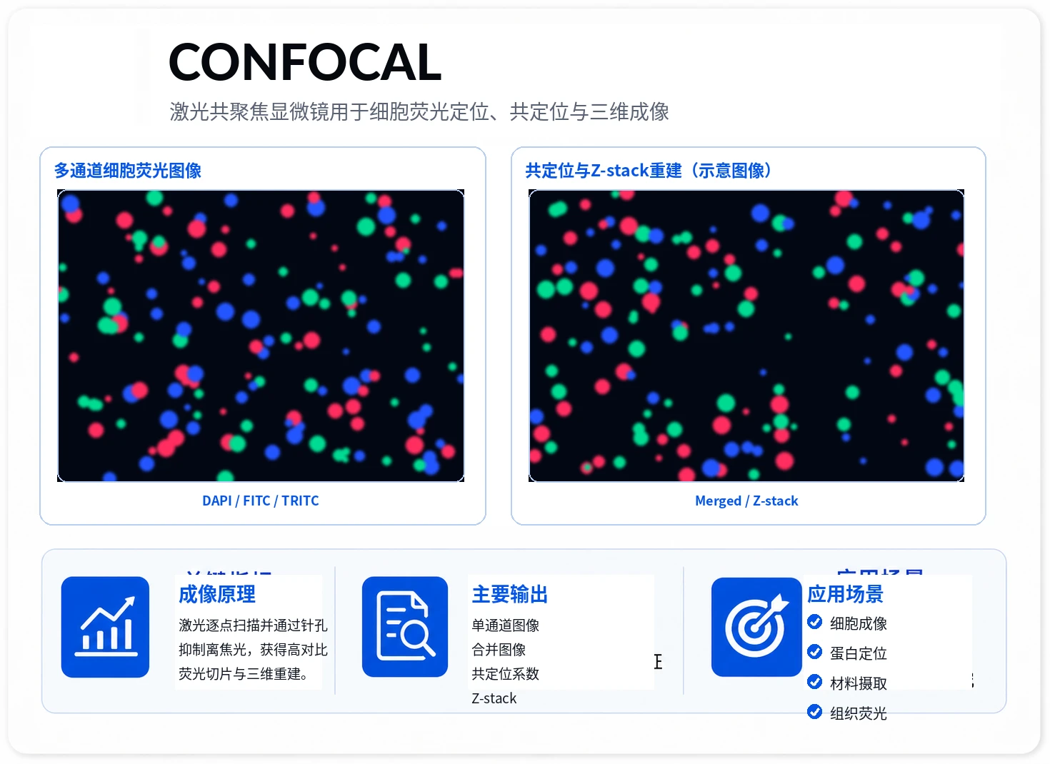
Cell fluorescent imaging
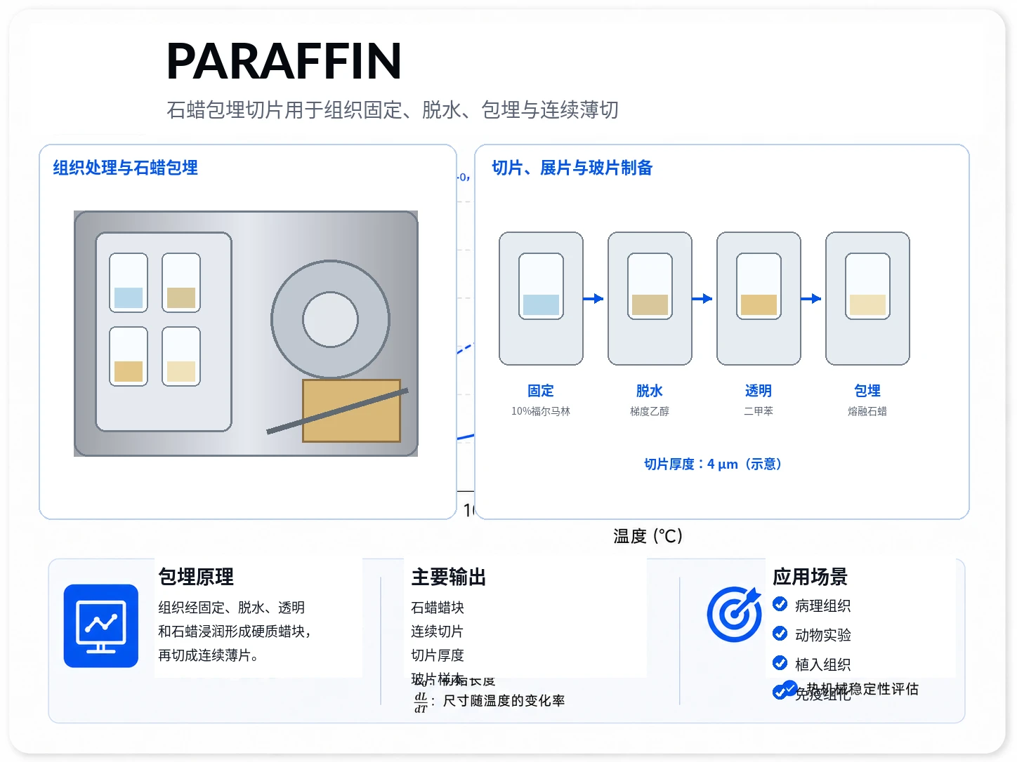
Division bag the slices.
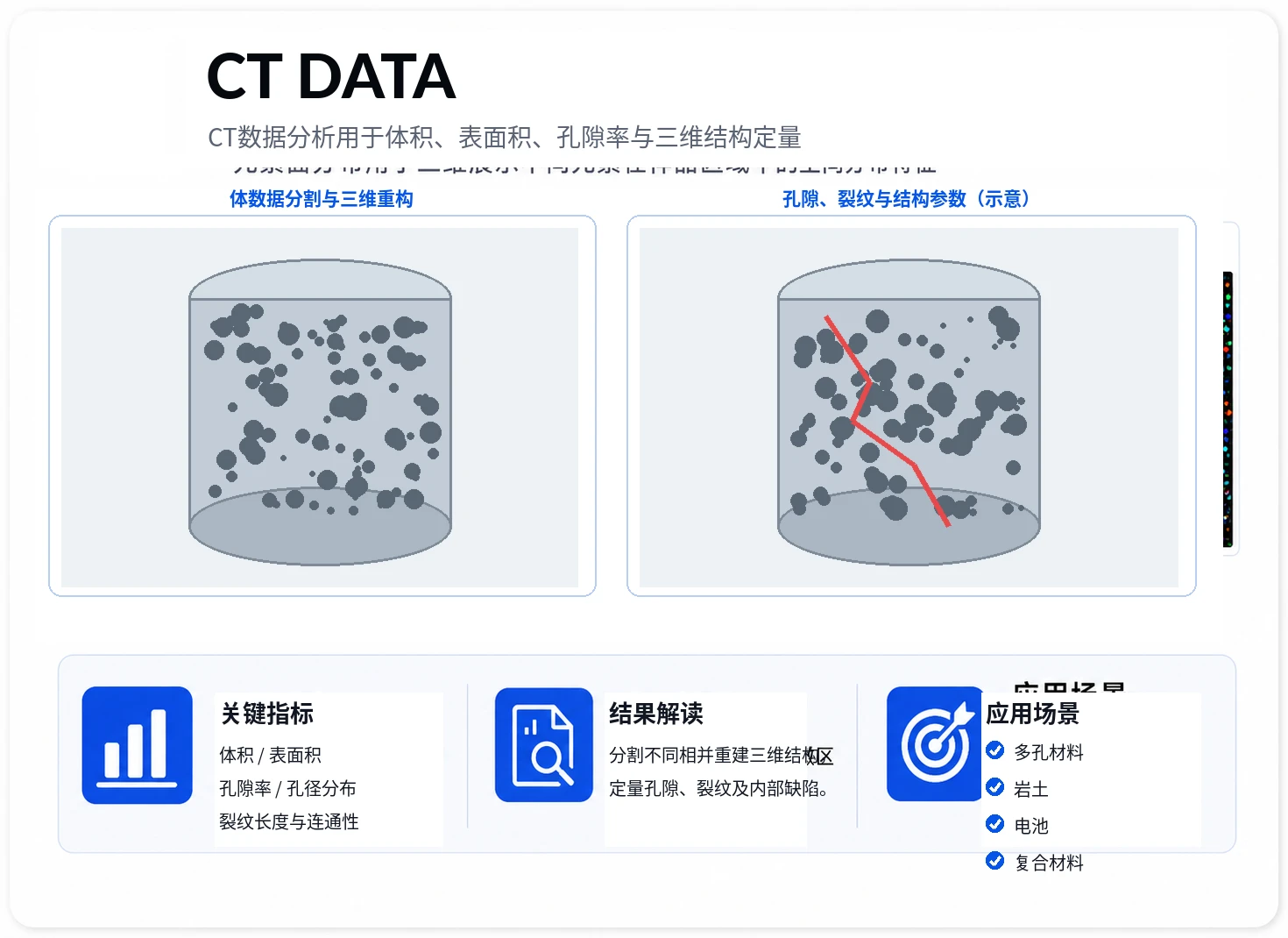
Volume, Table area, Peak Rate, Reconstruct 3D
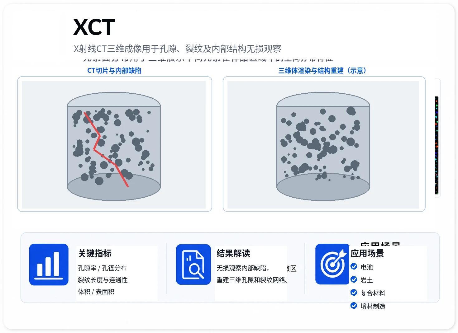
Hole/cleavage/internal structure
 AI / Simulation
AI projects, DFT, MD, AIMD, data analysis, and software development
 Characterization / Supplies
Characterization, supplies and instruments, contracts, invoices, shipping, and after-sales support
Applicable to AI project, DFT, MD, AIMD, data analysis, scientific mapping and software development needs. For addition, please comment on "OfficeNet Consulting + Unit + Demand Keywords".
For characterization, laboratory supplies and instruments, contracts, payments, invoices, shipping, and after-sales support.